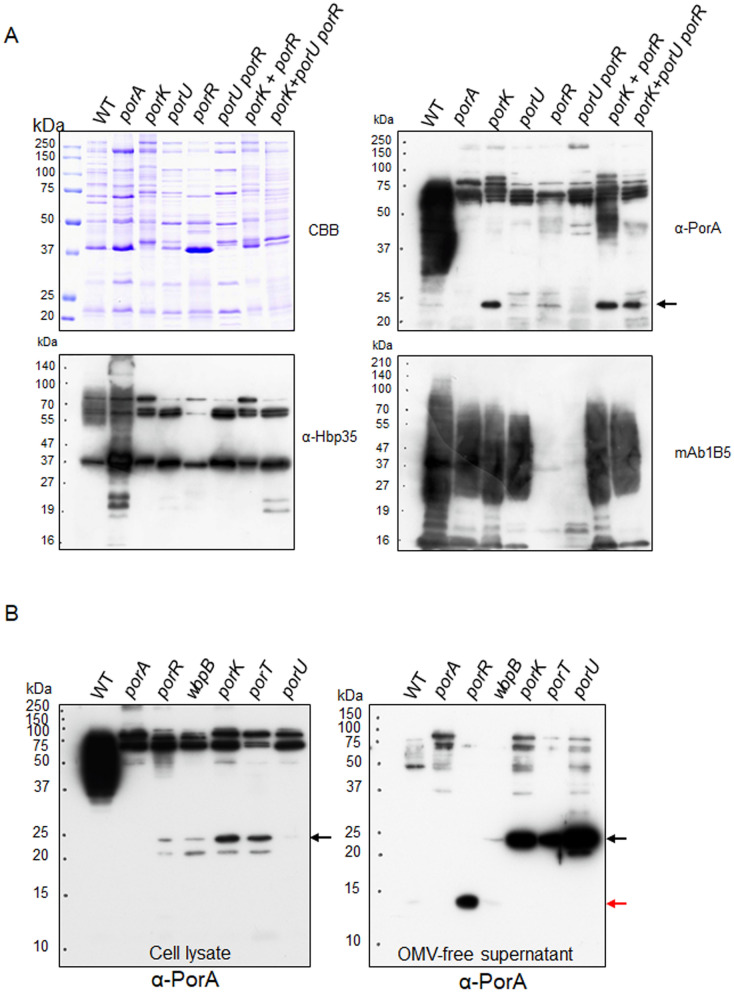
Figure 5.
Appearance of A-LPS-bound PorA in co-culture of the porK and porR mutants. (A) OMV fractions of cultures of the wild type, ΔporA, porK, porU, ΔporR, and porU ΔporR strains and co-cultures of the porK and ΔporR strains, and the porK and porU ΔporR strains were subjected to SDS-PAGE, followed by immunoblot analyses using α-PorA, α-Hbp35, and mAb 1B5. CBB: Coomassie Brilliant Blue staining. The black arrow indicates the 23-kDa CTD-containing PorA. (B) Cell lysates (left) and OMV-free culture supernatants (right) of the wild type, ΔporA, ΔporR, wbpB, porK, porT, and porU strains were subjected to SDS-PAGE, followed by immunoblot analysis with α-PorA, respectively. The black and red arrows indicate the 23-kDa CTD-containing PorA and the 14-kDa CTD-lacking PorA, respectively.

