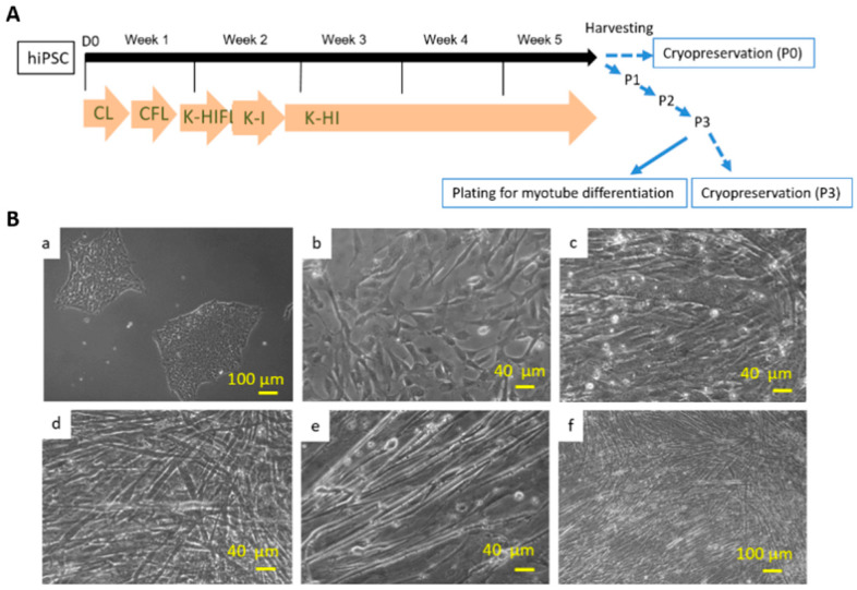
Figure 1.
(A) Diagram of the myogenic differentiation protocol from hiPSCs. The differentiation protocol from Week 1~5 is basically the same as in Chal’s publication. The culture was then harvested for cryopreservation or 3X series passaging as delineated by the blue arrows, which is the beneficial modification developed in this study. (B) Differentiation of myotubes from human iPSCs demonstrated by phase microscopy images. (a) iPSCs before myo-lineage differentiation. (b) myogenic progenitors before myotube induction. (c–f) Myotubes at different stages of differentiation and at different magnifications. (c) Day 4; (d) Day 7; (e,f) Day 8.

