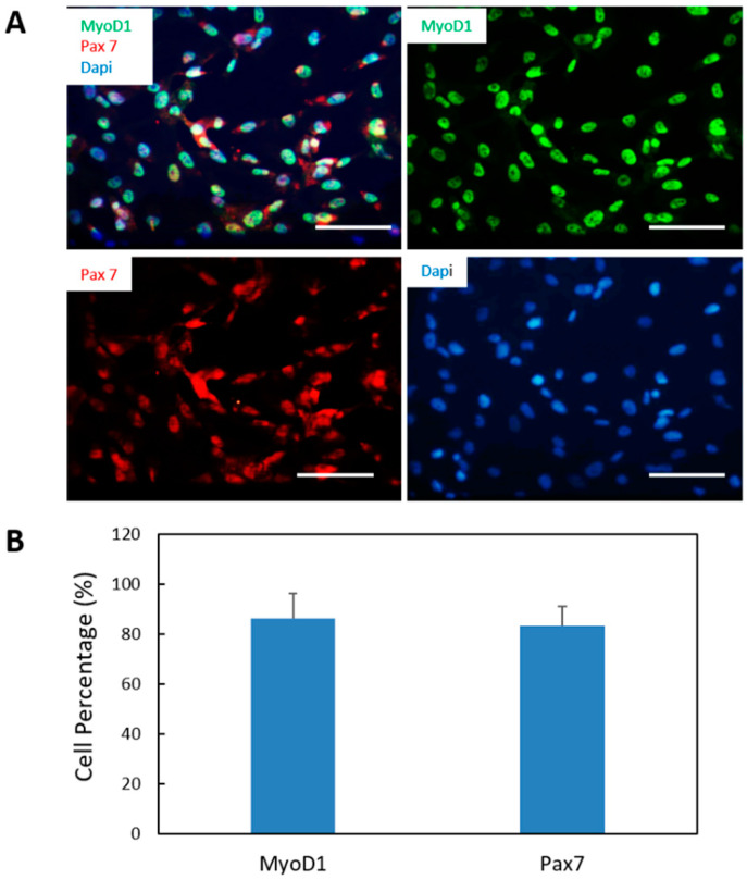
Figure 2.
Characterization of myogenic progenitors differentiated from iPSCs. (A) Myogenic progenitors differentiated from iPSCs were expanded and immunostained for the myoblast markers MyoD1 (green) and Pax 7 (red). (B) Quantification of the percentages of MyoD1-postive cells and Pax7-positive cells out of the total number of cells (visualized by DAPI based on the immunostaining from A. Over 15 images were randomly taken and analyzed for each coverslip. Three coverslips from three different batches of experiments were analyzed. Data presented are mean ± SEM. Scar bars: 100 µm.

