Figure 8.
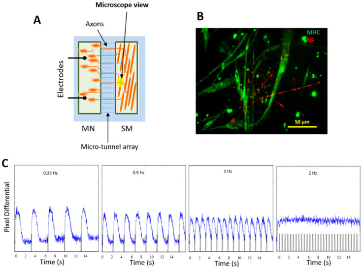
Innervation of iPSC-SKM by iPSC-MNs derived from the same iPSC line. (A) A diagram of the NMJ chamber (a courtesy from Santhanam et al. [18]). (B) iPSC-derived muscle culture was incorporated into the NMJ chamber system and co-cultured with motoneurons derived from the same iPSC line. Myotubes and MN axons in the muscle chamber were visualized in the muscle chamber through immunostaining utilizing antibodies against MHC (green) and Neurofilament (red) respectively. Analysis was done on Day 12 of the co-culture. The extensive distribution of axonal terminals in the muscle chamber and their physical interactions with myofibers was demonstrated by immunostaining with antibodies against Neurofilament and myosin heavy chain. (C) Innervated myofibers contracted in response to motoneuron stimulation under all frequencies tested.

