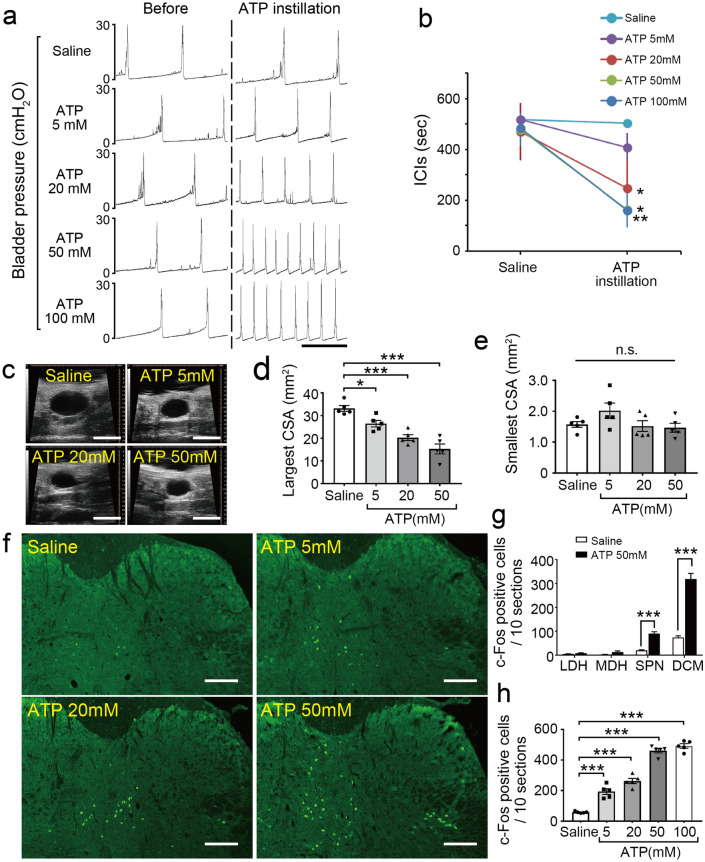
Figure 6.
Intravesical ATP treatment triggers activation of L6–S1 spinal neurons and induces an enhanced micturition reflex. (a) Representative cystometrograms after ATP instillation in wild-type mice. Scale bar, 10 min. (b) Changes in ICIs after ATP treatment (n = 5 mice per group); *p = 0.041 for 20 mM ATP, *p = 0.010 for 50 mM ATP, and **p = 0.0018 for 100 mM ATP (paired t-test with Holm correction). (c) Ultrasonographic findings of pre-voiding with intravesical ATP treatment. Scale bars, 5 mm. (d, e) Changes in largest CSA (d) and smallest CSA, post-voiding (e) (n = 5 mice per group); *p = 0.041 in saline versus 5 mM ATP, and ***p < 0.001 in saline versus 20 mM ATP and 50 mM ATP (Tukey’s test following one-way ANOVA). (f) Immunohistochemical analysis of c-Fos expression in L6–S1 spinal cord after ATP instillation. Scale bars, 100 μm. (g) The distribution of c-Fos-positive cells in the L6–S1 spinal cord induced by ATP instillation (50 mM) in wild-type mice (n = 5 mice per group); ***p < 0.001 in SPN and DCM (Student’s t-test with Holm correction). (h) The number of c-Fos-positive cells in L6–S1 spinal cord after ATP instillation (n = 5 mice per group: 10 sections per mouse were assessed). ***p < 0.001 in saline versus all ATP concentrations (Tukey’s test following one-way ANOVA). Error bars represent s.e.m., n.s., not significant.

