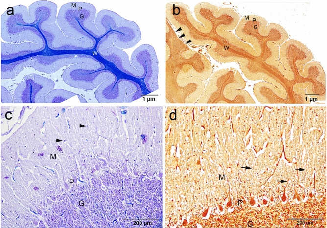
Figure 2.
Histochemical staining showing the general architecture of the camel cerebellum. (a) Cresyl violet with luxol fast blue, (b) silver staining showing the cerebellar medulla white matter (W) and three layers of the cerebellar cortex; M (molecular layer), P (Purkinje layer) and G (Granular layer). (b) Notably, a wide region of white matter that reach distally to the pial surface, without cortical tissue covering (arrowheads). (c) The outer molecular layer (M) consisted of relatively few numbers of neuronal cell bodies (arrowheads). (d) Silver staining showing more polymorphic neuronal cells in the molecular layer compared to cersyl violet staining. Moreover, different processes of numerous cells could be visualized by the silver stain (arrows).

