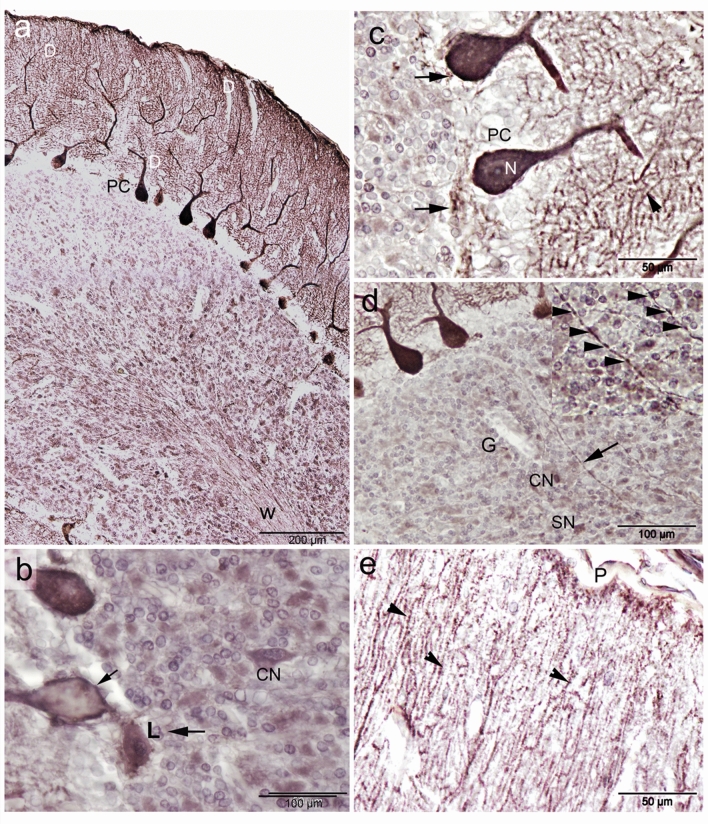
Figure 5.
Calbindin-D28k (CB) immunoreactivity in camel cerebellum. (a) CB immunoreactivity was obvious in almost all cerebellar cortical layers and the white matter (W). Purkinje cells (PC) showed the highest CB immunoreactivity. In addition, the dendrites extend the whole length of the molecular layer, showing an obvious dendritic arborization (D). (b) Seldom low CB-immunopositive PC bodies were observed (arrow). Neuron of Lugaro (L) and candelabrum neurons (CN) were observed. (c) CB immunoreactivity was seen as densely packed homogeneous deposits in the cytoplasm of the Purkinje cells and within the nuclei (N). Notice, CB immunoreactive fibers beneath and surrounding the PC, which are likely from basket and/or Lugaro cells (arrow). Immunoreactivity was observed in the spiny branchlets (arrowhead). (d) CB-immunopositive fibers (arrow) were traversing through the granular layer (G) and were oriented vertically till reaching the white matter. Inset showing the varicosities (arrowheads) along the axon profile which originated from the PC. (e) CB immunoreactivity was observed along the dendritic arborization till reaching the pial surface (P), arrowheads indicates the spiny branchlets of the dendrities.

