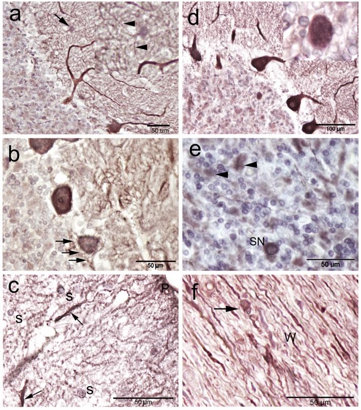
Figure 6.
Calbindin-D28k (CB) immunoreactivity in camel cerebellum. (a) Basket neuron (arrow) was recognized by their elongated body whose long axis (arrowheads) parallel to the cerebellar surface. (b) CB immunopositive rim surrounding the cell body surface of the Purkinje neurons (PC), which corresponding to axon terminals of the basket neurons (arrows). (c) The outer zone of the molecular layer showing the polymorphic stellate cells (S). Notice, the strong CB immunoreactivity was also observed within the molecular layer coming from the deeply stained dendrites of PC (arrows) which reaches to the distal ramifications just beneath the pial surface (P). (d, e) The granular cell layer showing a moderate CB immunoreactivity, however, the expression was heterogeneous. (d) An inset showing the Calbindin immunoreactivity by Golgi neurons. (e) Synaromatic (SN) large neurons were observed among the granular cell layer. A cellular spots or island in the granular layer (arrowheads) were observed. (f) The perivascular large non-traditional neurons (arrowheads) surrounding the blood vessels (BVs) showed positive immunoreactivity with CB in the cerebellar medulla or white matter (W).

