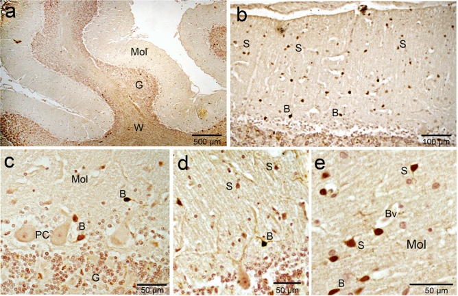
Figure 9.
Calretinin (CR) immunoreactivity in camel cerebellum. (a) Calretinin immunoreactivity was obvious in almost all cell layers with varying degrees. (b) The molecular layer cells (M) showed strong immunoreactivity for calretinin. (c, d) The basket neurons (B) were recognized by their elongated body whose long axis (arrowhead) parallel to the cerebellar surface and localized in the inner region of the molecular layer. Purkinje cells (PC) showed any or very weak immunoreactivity for Calretinin. (e): The stellate neurons (S) were characterized by their polymorphic body and their localization in the outer zone of the molecular layer. Mol molecular layer, BV blood vessel.

