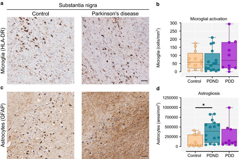
Fig. 3.
Microglial activation and astrogliosis in the substantia nigra. a Representative image of HLA-DR+ microglia in the substantia nigra of a control (left) and a Parkinson’s brain (right). The dark brown pigmented cells are neuromelanin-containing dopaminergic neurons. b Quantification of the total activated (enlarged amoeboid) microglia per mm2 (Kruskal–Wallis with Dunn’s multiple comparisons test, p = 0.646). c Representative image of astrocytic GFAP immunostaining in the substantia nigra of a control (left) and a Parkinson’s brain (right). d Quantification of the total GFAP-stained area per mm2 (Kruskal–Wallis with Dunn’s multiple comparisons test, p = 0.024; Control vs PDND p = 0.019). Control n = 12, PDND n = 16, PDD n = 10. PDND Parkinson’s disease no dementia, PDD Parkinson’s disease dementia. Scale bar: 100 μm. *p < 0.05

