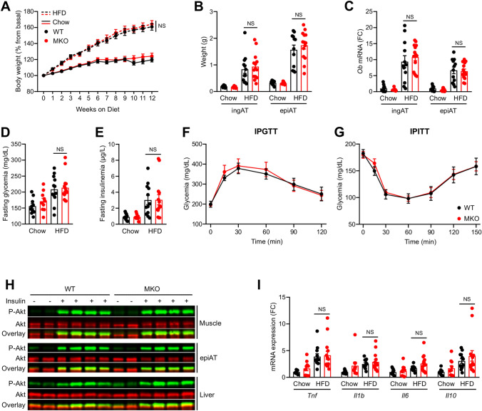
Figure 2.
Effect of RORα deletion in macrophages on HFD-induced obesity and IR. Ten weeks old WT and MKO mice were fed with either a chow or a HFD for 12 weeks. (A) Body weight gain. (B) Weight of inguinal (ingAT) and epidydimal (epiAT) adipose tissues. (C) mRNA expression levels measured by RT-qPCR for the Ob gene in both ingAT and epiAT. (D,F) After 10 weeks of diet, mice were fasted for 5 h and glycemia (D) and insulinemia (E) were measured, then intraperitonealy injected with 1 g/kg glucose for a glucose tolerance test (IPGTT) (F). (G) Intraperitoneal insulin tolerance test (IPITT) was performed by injecting 1 IU/kg insulin after 11 weeks of diet and 5 h of fasting. (H) Western blot of phospho-AKTSer473 in skeletal muscle, epiAT and liver. Mice were injected with either PBS or 1 IU of insulin 15 min before sacrifice. Western blots were scanned with an Odyssey CLx Imaging System (LI-COR) and with the Image studio Version 4.0.21 software (LI-COR, https://www.licor.com/bio/image-studio/). Images were cropped for sake of clarity and full-length blots are presented in Supplementary Figs. 7 to 9. (I) mRNA expression levels measured by RT-qPCR for Tnf, Il1b, Il6, and Il10 genes in epiAT. Data are shown as mean ± SEM. 2-way ANOVA followed by Sidak’s multiple comparisons test was performed. All statistical analyses were carried out using GraphPad Prism 8 for Windows (GraphPad Software). n = 10–15 mice per group. NS: Not significant; FC: Fold Change. See also Fig.S2.

