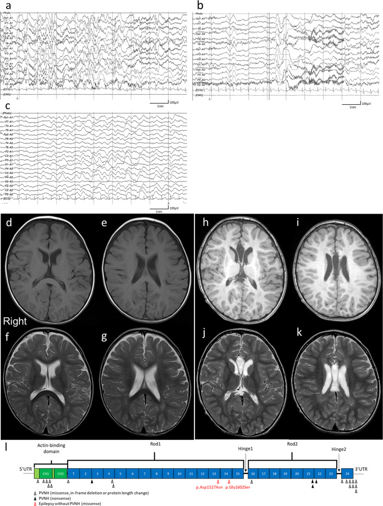
Fig. 1. Clinical information of the patient and structure of filamin A protein.
a Electroencephalography (EEG) results of the patient showing hypsarrhythmia during wakefulness at 9 months of age. b Ictal EEG results showing cluster spasms with head drop at 9 months of age. c EEG results at 11 months of age and after adrenocorticotropic hormone therapy. Sporadic slow waves were observed in the bitemporal regions. d, e Brain MRI T1-weighted and f, g T2-weighted images of the patient at the age of 15 months. Three pediatric neurologists independently confirmed the absence of periventricular nodular heterotopia. h, i Brain MRI T1-weighted and j, k T2-weighted images of the patient at the age of 29 months. l Filamin A protein with functional domains and FLNA variants found in males. Functional domains consist of the actin-binding domain containing two calponin homology (CH) domains and 24 Ig domains. Pathogenic variants detected in males are shown as triangles below Filamin A. FLNA variants in PVNH1 or in epilepsy without PVNH are shown as black or red triangles. Filled or open triangles indicate nonsense or missense/in-flame changes.

