
Figure 1.
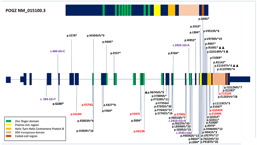
Schematic representation of the POGZ protein and POGZ gene. Pathogenic variants in the present study shown above the exon structure and variants previously reported in the literature shown below. Black font—frameshift or nonsense variants. Purple font—splice variants. Red font—missense variants. Triangles mark additional reports of the same variant