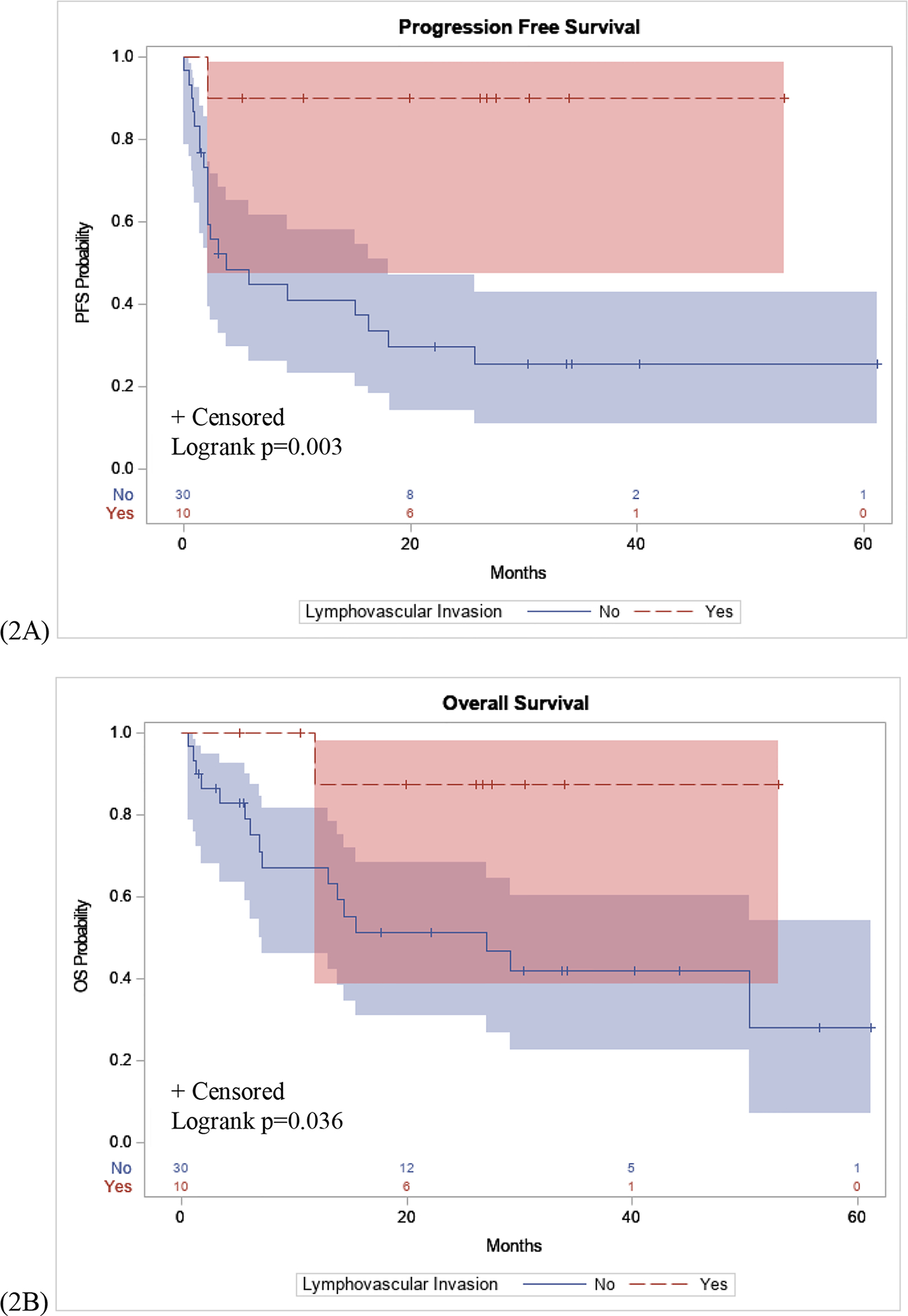
Figure 2: PFS and OS Stratified by Lymphovascular Invasion.

(2A): Patients with lymphovascular invasion seen on primary biopsy samples had a significantly longer PFS interval following immunotherapy compared to patients without lymphovascular invasion (p=0.003). (2B): Significant difference in OS in patients with lymphovascular invasion compared to patients without lymphovascular invasion (p=0.036).
