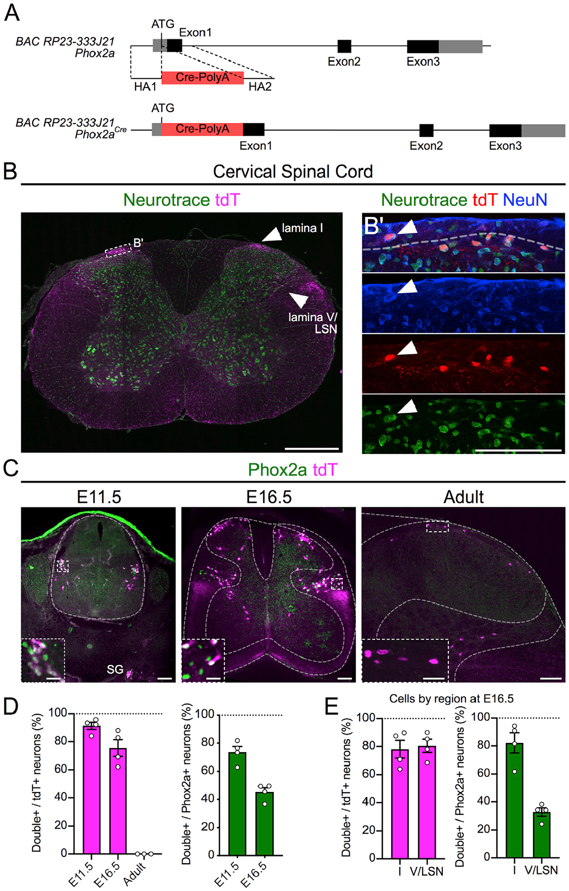
Figure 1. Spinal Phox2aCre Neurons Reside in Lamina I, Lamina V, and LSN All images are of Phox2aCre; R26LSL-tdT/+ mice.

(A) BAC recombination strategy: Cre-PolyA insertion 3′ to the Phox2a ATG codon in the BAC RP23–333J21.
(B) tdTomato (tdT)+ neurons in lamina I, lamina V, and LSN of the cervical spinal cord of adult mice.
(B’) Magnified box in (B) showing lamina I Neurotrace, tdT, and NeuN co-labeling.
(C) Expression of tdT and Phox2a in E11.5, E16.5, and adult mouse spinal cord.
(D) Percentage of tdT+ neurons that express Phox2a, as well as percentage of Phox2a+ neurons that express tdT at E11.5 and E16.5 and in adult mice.
(E) Percentage of tdT+ neurons that express Phox2a, as well as percentage of Phox2a+ neurons that express tdT in the superficial and deep dorsal horn of E16.5 mouse spinal cords.
n = 4 E11.5 mice, n = 4 E16.5 mice, and n = 3 adult Phox2aCre; R26LSL-tdT/+ mice. Data are represented as mean ± SEM. Scale bars: 500 μm in (B), 100 μm in (B’), 100 μm in (C), and 25 μm in insets. SG, sympathetic ganglia.
