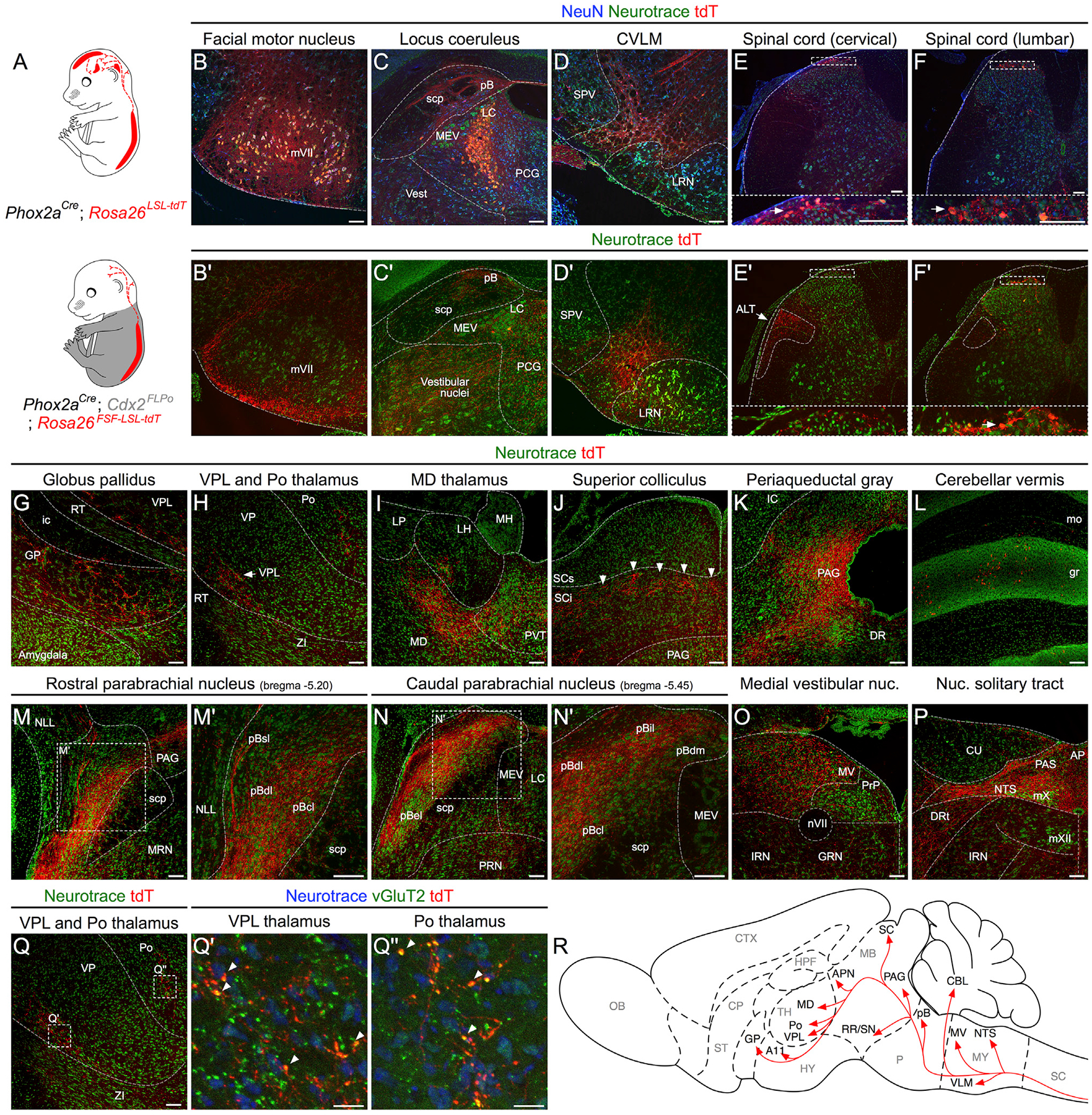
Figure 2. Spinal Phox2aCre Neurons Innervate AS Targets.

(A) Intersectional genetic strategy to visualize spinofugal axons with tdT. Phox2aCre; R26LSL-tdT/+ mice have tdT cellular expression in the brain and spinal cord, while Phox2aCre; Cdx2FlpO; R26FSF-LSL-tdT/+ mice have cellular tdT expression only in spinal Phox2a neurons.
(B–F’) In (B)–(F): expression of cellular tdT in the brain and spinal cord of Phox2aCre; R26LSL-tdT/+ mice in comparison to (B’–F’) the lack of tdT expression in the brain and spinal cord of Phox2aCre; Cdx2FlpO; R26FSF-LSL-tdT/+ mice, except caudal to the cervical level. In (E’), arrow indicates presumptive anterolateral tract (ALT) axons in white matter. Insets in (E), (F), (E’), and (F’) correspond to stippled boxes and show tdT+ cell bodies (white arrows)
(G–P) Prominent targets of tdT+ spinofugal axons. Higher magnifications are shown in (M’) and (N’).
(Q–Q”) vGluT2 and tdT immunohistochemistry in the thalamus demonstrates putative excitatory synaptic termini arising from spinofugal axons. The image in (Q) is duplicated from (H) and is used as a reference for (Q’) and (Q”).
(R) Diagram summarizing the termination sites of tdT+ spinofugal axons.
n = 3 Phox2aCre; R26LSL-tdT/+ adult mice, and n = 3 Phox2aCre; Cdx2FlpO; R26FSF-LSL-tdT/+ adult mice. Scale bars: 100 μm, except 25 μm for (Q’) and (Q”). AP, area postrema; CU, cuneate nucleus; DR, dorsal raphe; DRt, dorsal reticular nucleus; GRN, gigantocellular reticular nucleus; ic, internal capsule; IRN, intermediate reticular nucleus; LH, lateral habenula; LP, lateral posterior thalamus; LRN, lateral reticular nucleus; MEV, midbrain trigeminal nucleus; MH, medial habenula; mo, molecular layer of the cerebellum; MV, medial vestibular nucleus; mVII, facial motor nucleus; mX, vagal motor nucleus; mXII, hypoglossal motor nucleus; NLL, nucleus of the lateral lemniscus; nVII, facial motor nerve; PAS, parasolitary nucleus; pBdm, dorsal-medial parabrachial nucleus; PCG, pontine central gray; PRN, pontine reticular nucleus; PRP, nucleus prepositus; PVT, paraventricular thalamus; RT, reticular thalamic nucleus; SCi, superior colliculus, intermediate laminae; scp, superior cerebellar peduncle; SCs, superior colliculus, superficial laminae; SPV, spinal trigeminal nucleus; ZI, zona incerta.
