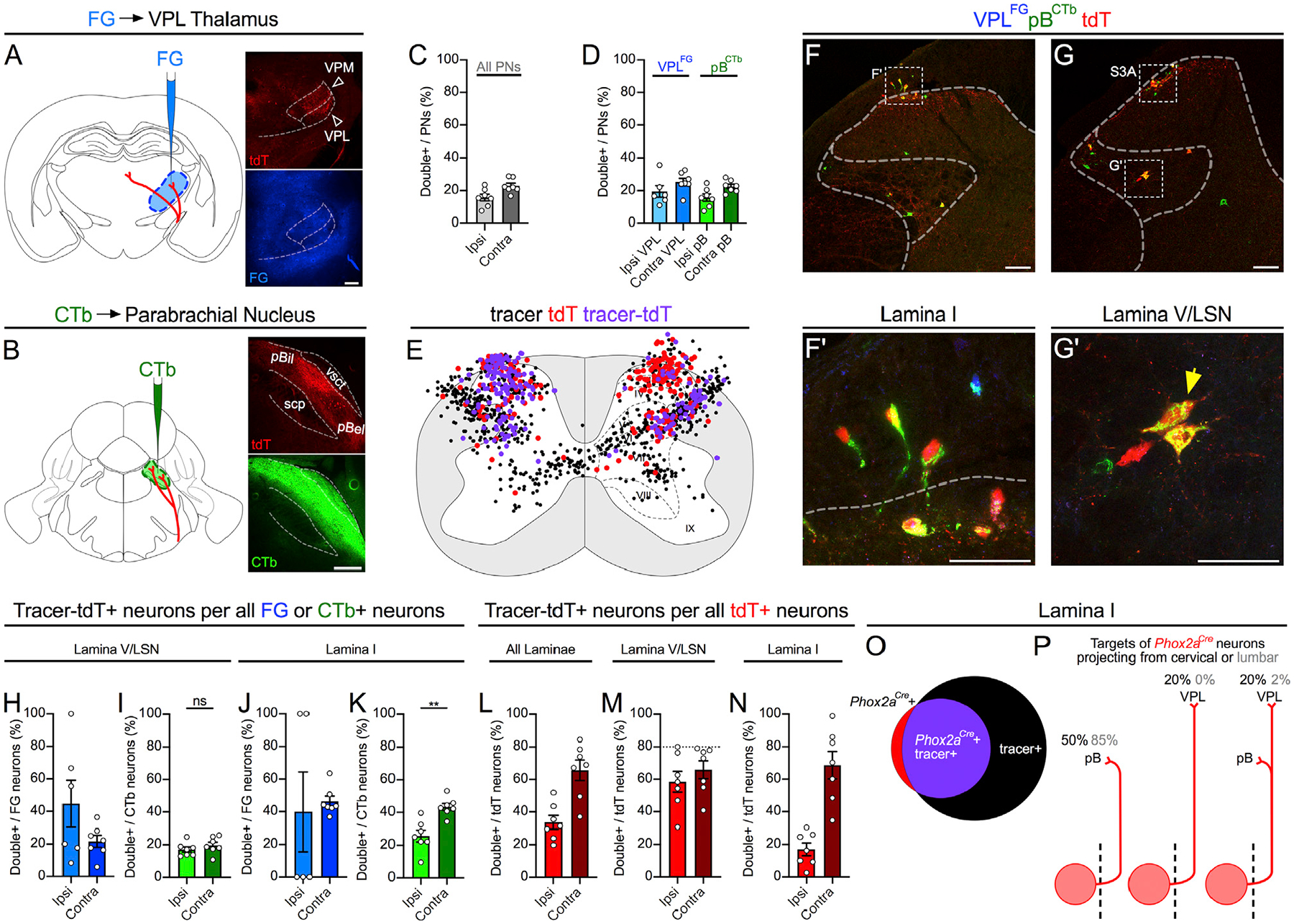
Figure 3. Spinal Phox2aCre Neurons Are Predominantly AS Neurons.

(A and B) Adult Phox2aCre; R26LSL-tdT/+ mice were injected with FG in the VPL thalamus (A) and CTb-488 in the parabrachial nucleus (B).
(C and D) Percentage of cervical spinal cord dorsal horn projection neurons expressing tdT, classified as those labeled with either tracer (C; All PNs) or selectively with FG or CTb (D).
(E) Diagram of the locations of tdT+ only, retrograde-labeled only (FG or CTb), or tdT+ and tracer-labeled (violet) neurons, in 5 non-sequential 25-μm sections of the cervical spinal cord of one representative animal.
(F–G’) In (F) and (G): representative images of the cervical spinal cord demonstrating tdT+ neuron labeling by retrograde tracers. See Figures S3A and S3B for more examples. (F’ and G’) High magnification of boxed areas in (F) and (G), depicting retrograde-labeled lamina I (F’) or lamina V/LSN (G’) tdT+ neurons (indicated by yellow arrows).
(H–N) Laminar analysis of neuron location in the cervical spinal cord ipsilateral or contralateral to tracer injection.
(H–K) Percentage of tracer-labeled neurons in lamina I (H and I) or lamina V/LSN (J and K) also expressing tdT, separated by tracer type.
(L–N) Percentage of tdT-labeled neurons labeled with one or both tracers in all laminae (L), in lamina V/LSN (M), or in lamina I (N).
(O) Diagram depicting overlap between tdT and retrograde tracer in lamina I of a representative mouse.
(P) Diagrams illustrating the estimated percentages of cervical and lumbar lamina I Phox2aCre neurons projecting to mouse pB, VPL, or both. Stippled line represents spinal midline.
n = 7 Phox2aCre; R26LSL-tdT/+ adult mice (4 male, 3 female). Mann-Whitney test in (I) and (K); **p < 0.01; ns, non-significant. Data are represented as mean ± SEM. Scale bars: 250 μm in (A) and (B), 100 μm in (F) and (G), and 50 μm in (F’) and (G’).
