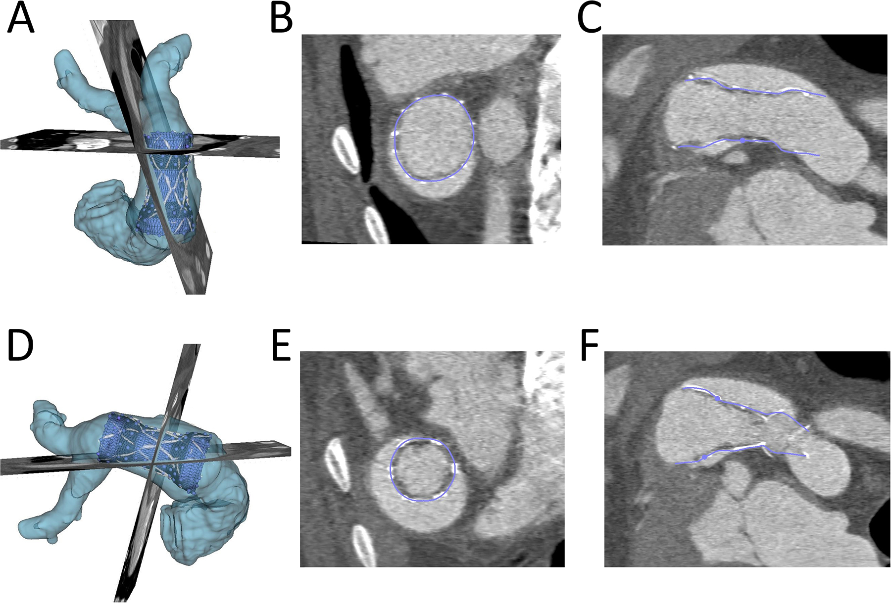
Figure 3. Demonstration of Ability of Virtual Device to Model Actual Device Placement in Post-Implant CT Scan:

The actual volume rendered device (white), RVOT(light blue), the conformed virtual device (dark blue) are shown. A) and D): Device conformation in 3D from anterior and lateral view; B) and E) Device conformation in two axial planes; C) and F) Device conformation in two sagittal planes.
