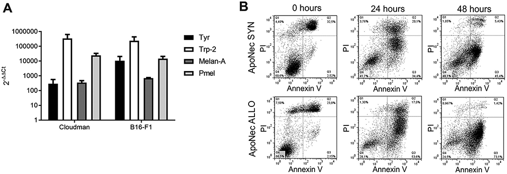
Fig. 1.

MDA expression and apoptosis induction after γ-irradiation. (A) mRNA levels for MDAs were assessed by qRT-PCR in B16-F1 and Cloudman cells. The 2−ΔΔCt values were calculated using B16-F1 and Cloudman Cts compared to H5V endothelial cells Cts. Actb was used as reference gene. Mean ± SD from two independent experiments is shown. (Studenťs t test, p > 0.05). (B) B16-F1 and Cloudman cells were γ-irradiated (70 Gy) and cultured for 0, 24 or 48 h. They were then stained with AnV-FITC and IP, and percentages of apoptotic/necrotic cells were assessed by flow cytometry.
