Fig. 2.
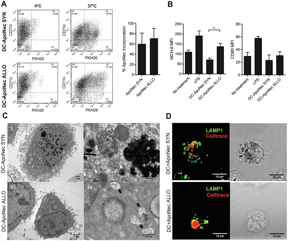
Internalization of ApoNec cells by DCs. (A) ApoNec SYN or ApoNec ALLO cells were stained with PKH26 and cultured with DCs for 24 h at 37 °C or 4 °C (unspecific binding). DCs were stained with anti-CD11c Ab, and PKH26 incorporation in CD11c+ cells was assessed by flow cytometry. Three independent experiments were performed, dotplots from a representative experiment are shown. The percentage of incorporation of ApoNec cells by DCs was assessed as the percentage of CD11c+ PKH26+ cells/CD11c+ cells. Mean ± SD from three independent experiments is shown. (Studenťs t test, α = 0.05, n = 3, p > 0.05). (B) MHC-II and CD86 MFI on DC-ApoNec was assessed by staining with anti-CD11c Ab and either anti-I-Ab or anti-CD86 antibody and analyzed by flow cytometry. Lipopolysaccharide (LPS)-treated DCs were used as positive control. Student’s t test was used to compare DC-ApoNec SYN and DC-ApoNec ALLO (α = 0.05, n = 3, *p < 0.05). (C) Transmission electron microscopy of DC-ApoNec SYN and DC-ApoNec ALLO (i) DC showing membrane ruffles and multiple endocytic/phagocytic compartments, some loaded with pigment (7000×). (ii) Endocytic/phagocytic compartments containing pigment granules (white arrows) shown at higher magnification (50,000×). (iii) DC showing membrane ruffles and endocytic/phagocytic compartments (12,000×). (iv) Macropinosomes (black arrows) shown at higher magnification (50,000×). (D) DCs were cultured for 6 hs with Celltrace violet-stained ApoNec SYN or ApoNec ALLO (red). Then, lysosomes in DCs were stained using anti-Lamp-1 Ab (green) and visualized using confocal microscopy.

