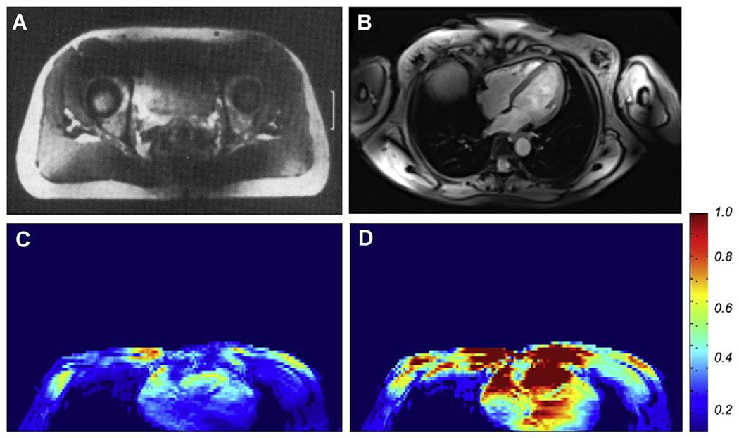
Fig. 4.

(A) Single axial slice image from the human pelvis reported from early 4 T experiments from the research laboratories of Siemens; (B), a contemporary 7 T image of an axial slice in the human torso, targeting imaging of the heart, obtained with a 16-channel transmit and receive array coil, using B1 “shimming”. (C) and (D) The transmit B1 magnitude map before (C) and after (D) optimization over the heart in an axial slice approximately at the same position as that shown in Fig. 4B, demonstrating that the B1 is normally highly inhomogeneous and weak over this organ of interest (C) but can be improved significantly by multichannel transmit methods (D).
