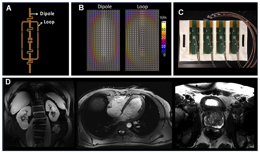
Fig. 6.

(A) The loop and dipole element within a single block of the 16LD coil used to generate the images in Fig.5C; (B) the electrical fields associated with the dipole and the loop elements, calculated by electromagnetic simulations. (C) Four loop-dipole blocks as manufactured containing the 8 transceiver channels for the anterior side of the 16LD coil. The same configuration is then placed also on the posterior side, as shown in Fig. 7E with a torso phantom. (D) Kidney, cardiac, and prostate images obtained with the 16LD coil.
