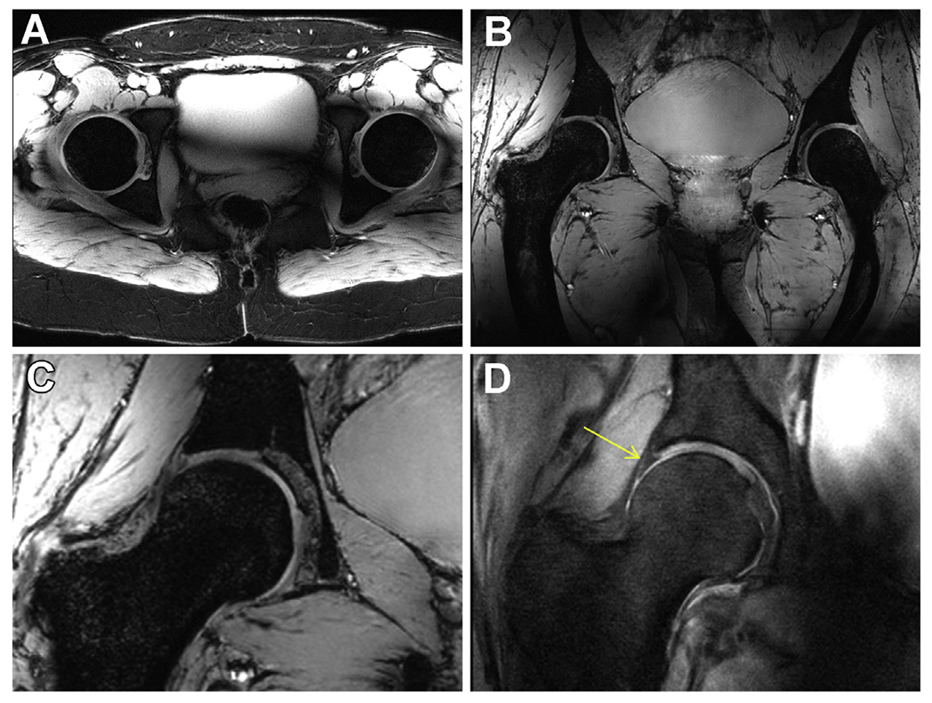
Fig. 9.

Anatomic hip imaging acquired at 10.5 T with a 10-channel dipole transceiver array. (A) An axial multislice 2D gradient echo acquisition acquired with fat saturation. (B) 3D coronal MEDIC acquisition. (C) Zoomed version of the right femoral head form the MEDIC (B) acquisition. (D) A proton-density (PD) weighted turbo spin echo (TSE) acquisition showing the expected contrast between the labrum and cartilage (yellow arrow).
