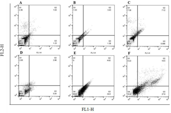
Fig. 2.
The effects of GSE on cell apoptosis was determined by flow cytometry. Treatment with GSE at 71 μg/mL for 24 and 48 h, significantly induced apoptosis in OVCAR-3 cells compared with the DMSO treated cells. (A and B) Control, cells without treatment with any substance in 24 and 48h respectively; (C and D) cells treated with DMSO as solvent of GSE for 24 and 48 h, respectively; and (E and F) cells treated with GSE at 71 μg/mL for 24 and 48 h, respectively. P < 0.001 indicates significant differences compared to the corresponding DMSO group GSE, Grape seed extract; DMSO, dimethyl sulfoxide

