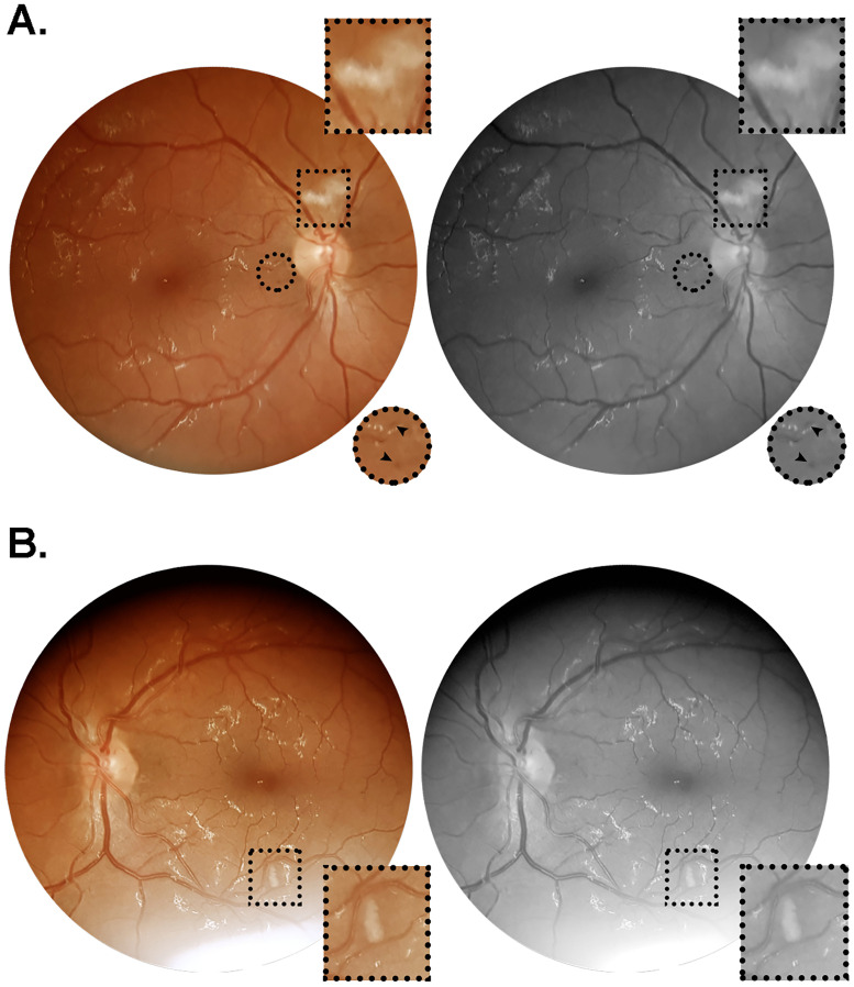
Fig 1. Color and red-free retinal photographs from patient #6 (W.A.C.).
Images acquired on day 64 after symptoms onset, from a 35 year-old obese and hypertensive man who was admitted on day seven since onset of fever, cough and dyspnea. During hospitalization, this patient evolved to critical illness, characterized by acute respiratory distress syndrome, respiratory failure and acute kidney injury, requiring ICU monitorization, mechanical ventilation and hemodialysis. The patient had a positive clinical outcome, and received hospital discharge on day 69 after symptoms onset. (A) Right retina showing a nerve fiber layer infarct above the optic nerve head, and microhemorrhages in the papillomacular bundle close to the optic disc. (B) Left retina showing nerve fiber layer infarcts at the inferior temporal vascular arcade, approximately 1.5 disc diameters inferior to the macula. Square and circular insets represent 2x magnification of the delimited area.

