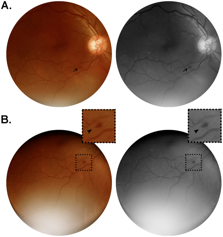
Fig 2. Color and red-free retinal photographs from patient #18 (W.S.).
Images acquired on day 16 from symptoms onset, from a 56 year-old man with no comorbidities, admitted on day 13 after onset of a clinical scenario characterized by fever, cough, headache, myalgia and dyspnea. The patient evolved to severe illness, not requiring ICU monitorization nor developing any organ failure. Lung CT at admission revealed bilateral ground-glass opacities predominantly in the periphery, extending to approximately 50% of lung parenchyma. (A) Flame-shaped hemorrhage (arrow) approximately one disc diameter from the optic nerve head, close to the inferior temporal vascular arcade. (B) Same lesion observed from an alternative point of gaze fixation, magnified in the insets (2x; arrowheads).

