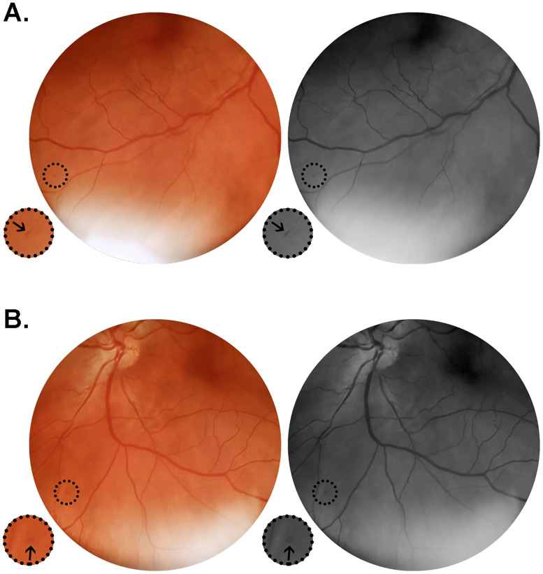
Fig 3. Color and red-free retinal photographs from patient #19 (G.C.X.).
Images acquired on day 12 from symptoms onset, from a 49 year-old man in regular treatment for arterial hypertension, admitted on the eight day after onset of fever, cough, myalgia and progressive dyspnea. Lung CT on admission showed typical viral pneumonia findings extending through 25–50% of the parenchyma. The patient evolved to a severe phenotype of the disease. (A) Right retina presenting with isolated microhemorrhage at the periphery close to the inferior temporal vasculature. (B) Left retina showing isolated microhemorrhage at the inferior nasal retinal quadrant. Insets represent delimited areas with 2x magnification.

