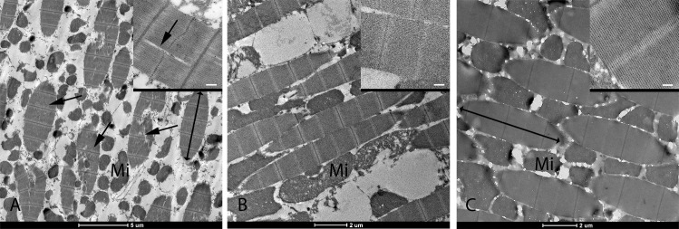
Fig 2. Transmission electron microscope images of the indirect flight muscles of Ae. aegypti female adults arranged within the thorax.
Filaments in the myofibrils appear to be disrupted in AeAct4hdr1 homozygotes (A, arrows and inset) but not in AeAct4hdr1 heterozygotes (B, inset) or WT (C, inset). Individual myofibrils are indicated with double-headed arrows, and mitochondria (Mi) in the muscle fiber cells are clearly visible. Insert scale bars = 200 nm.

