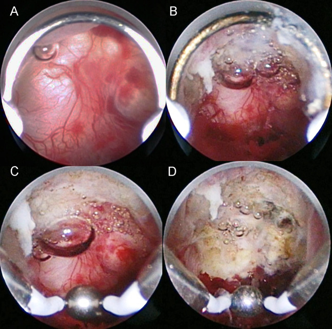
Fig 1. Intraoperative images of the hysteroscopic surgery procedure.
(A) Abnormal hypervascularity is observed in the cesarean scar defect. (B) Cutting of the inferior edge of the cesarean scar defect using a cutting loop electrode. (C) Cauterization of all areas including the abnormal vasculature in the cesarean scar defect. (D) Appearance after cauterization using a ball electrode.

