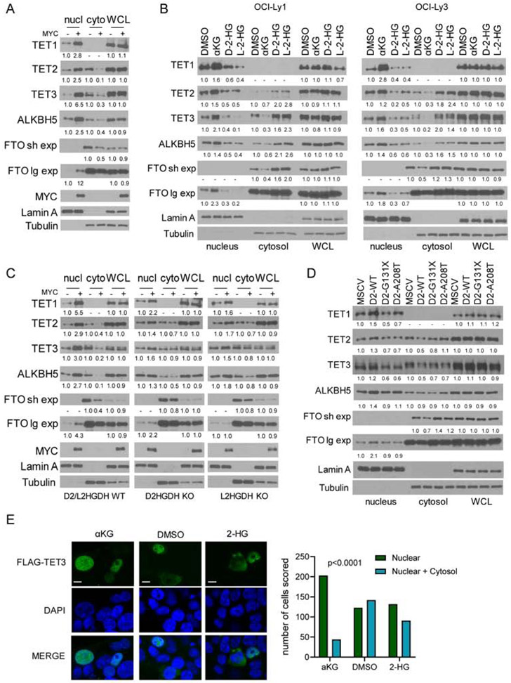
Figure 5. Control of the subcellular localization of TET1-3, FTO and ALKBH5 by MYC, D2HGDH/L2HGDH, and αKG/2-HG.
a) WBs of TET1-3, FTO and ALKBH5 in nuclear and cytoplasmic fractions, and whole cell lysate (WCL) of P493-6 cells upon MYC suppression and its re-expression. Two exposures (short and long) are shown for FTO. b) WBs of TET1-3, FTO and ALKBH5 in the subcellular fractions and WCL of DLBCL cell lines exposed to DMαKG (5mM), octyl-D-2-HG or octyl-L-2-HG (100μM) for 8h. c) WBs of TET1-3, FTO and ALKBH5 in subcellular fractions and WCL of P493-6 cells (D2HGDH and L2HGDH WT or KO) upon MYC suppression and its re-expression. d) WBs of TET1-3, FTO and ALKBH5 in HEK-293 cells expressing an empty vector (MSCV), D2HGDH WT or the G131X and A208T mutants. Densitometric measurements of protein levels are shown at the bottom of WB displays – all values are relative to controls and already corrected by WCL abundance, which is shown for reference. e) immunofluorescence of HEK-293T cells transiently transfected with a FLAG-TET3 plasmid and exposed to DMαKG (5mM), octyl-D-2-HG and L-2-HG (100μM/each) or DMSO for 6h; scale bar is 10 μm. Scoring of nuclear vs. nuclear and cytoplasmic expression in the three conditions is shown on the right; p<0.001, Chi-square.

