. 2020 Oct 21;9:e59882. doi: 10.7554/eLife.59882

Figure 5. CCMB1:pCB’+pCCM’ produces carboxysomes when grown in air.

(A) Polyhedral bodies resembling carboxysomes are evident in electron micrographs of CCMB1:pCB’+pCCM’ cells grown in air (full CCM, both images on the right) but were not observed in a negative control lacking pCB and pCCM plasmids (left, Methods). All panels have equal scale. (B) Carboxysome structures purified from CCMB1:pCB’+pCCM’ grown in ambient air (Materials and methods, right) resemble structures isolated from the native host (left) in size and morphology. Figure 5—figure supplement 2 gives full size and additional images clearly showing rubisco inside isolated carboxysomes. SDS-PAGE gels in Figure 5—figure supplement 1 demonstrate co-migration of rubisco large and small subunits with carboxysomes structures through the purification procedure.

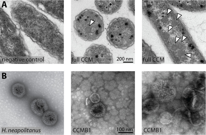
Figure 5.

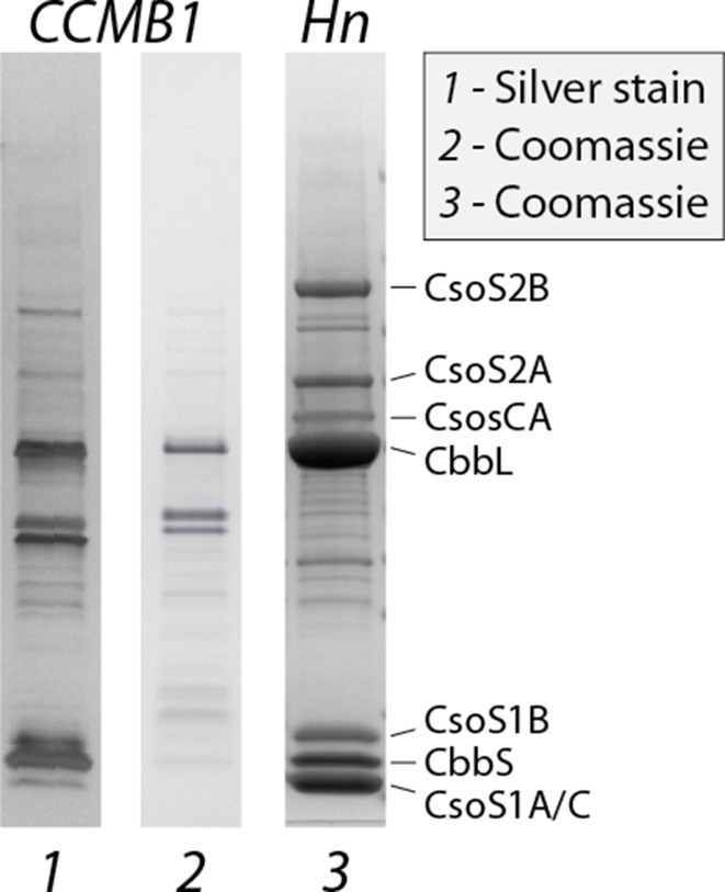
Figure 5—figure supplement 1. Carboxysomes purified from CCMB1:pCB’ + pCCM’ contain rubisco and other known carboxysome components.

Figure 5—figure supplement 1.

Lanes 1 and 2 give purifications from CCMB1:pCB’+pCCM’ grown in ambient air stained with silver and coomassie stains, respectively. Lane 3 is a purification from wild-type H. neapolitanus (Materials and methods). The legend on the right marks the carboxysome components that are typically visible on preparations from the native host: the shell proteins CsoS1CAB, two forms of the disordered protein CsoS2, the carbonic anhydrase CsoSCA, and the rubisco large and small subunits CbbLS. CbbLS bands are evident in all lanes, and all three purifications were found to contain carboxysome-like structures when imaged by transmission electron microscopy, as shown in Figure 5 and Figure 5—figure supplement 2.

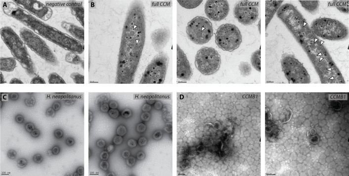
Figure 5—figure supplement 2. CCMB1:pCB’ + pCCM’ produces polyhedral bodies resembling carboxysomes when grown in ambient air.

Figure 5—figure supplement 2.

Thin section transmission electron micrographs of a negative control strain (A) and experimental cells (B) show that air-grown CCMB1:pCB’+pCCM’ cells (‘full CCM’ in panel B) contain morphological carboxysomes (white arrows). The negative control for carboxysome expression is CAfree:pFE-sfGFP + pFA-HCAII (Materials and methods). Expression of CCM was associated with production of black-staining stress granules, which were not observed in images of the negative control. (C) Carboxysomes isolated from wild-type H. neapolitanus displayed regular pseudo-icosahedral structures with ≈100 nm diameter, as expected (Shively et al., 1973). (D) Carboxysomes isolated from CCMB1 were less regular, but clearly resemble native structures. Preparations also contained ‘rosette’ structures we often observe when isolating carboxysomes from E. coli (far right panel). Purification yields from CCMB1:pCB’+pCCM’ were much lower than is typical for preparations from wild-type E. coli which may explain the relative abundance of rosette structures.