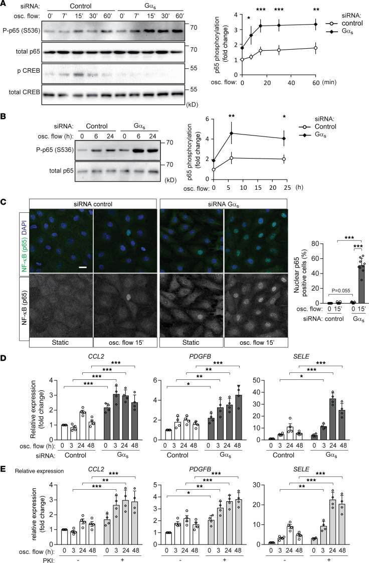
Figure 1. Knockdown of Gαs increases endothelial inflammation induced by disturbed flow in BAECs.
(A–D) Confluent BAECs were transfected with control siRNA or siRNA directed against Gαs and were then exposed to oscillatory (osc.) flow for the indicated time period. p65 and/or CREB phosphorylation was analyzed by immunoblotting (A and B, n = 3 independent experiments), cellular p65 localization was determined by staining of cells with an anti-p65 antibody (C, n = 3 independent experiments, at least 3 view fields were analyzed per experiment), and inflammatory gene expression was analyzed by qPCR (D, n = 4 independent experiments). The diagrams show the densitometric evaluation of p65 phosphorylation (A and B) or of nuclear p65 staining (C). Scale bar: 20 μm. (E) Confluent BAECs were incubated without or with the PKA-inhibitor PKI (1 μM). Inflammatory gene expression after osc. flow induction was analyzed (n = 4 independent experiments). Data represent mean values ± SD; *P ≤ 0.05, **P ≤ 0.01, ***P ≤ 0.001 (2-way ANOVA and Bonferroni’s post hoc test [A, B, D, and E] and 2-tailed Student’s t test [C]).

