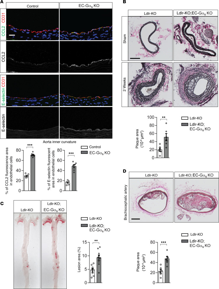
Figure 3. Induced loss of endothelial Gαs results in increased endothelial inflammation and atherosclerotic plaque formation.
(A) Cross-sections of the inner curvatures of aortic arches of control or EC-Gαs–KO mice stained with DAPI (blue) and antibodies against CCL2 or E-selectin (green) and CD31 (red). Bar diagrams show percentage of area stained by anti-CCL2 or anti–E-selectin antibodies of total endothelial cell area defined by anti-CD31 antibody staining (n = 6 animals per group; at least 3 sections were analyzed per animal). Scale bar: 25 μm. (B) Atherosclerosis-prone Ldlr-KO mice without (Ldlr-KO) or with endothelium-specific Gαs deficiency (Ldlr-KO;EC-Gαs–KO) were sham operated or underwent partial carotid artery ligation. Two weeks after the ligation, serial sections were made through the entire carotid arteries and stained with elastic stain (n = 6 mice per group). Scale bar: 100 μm. (C and D) Ldlr-KO or Ldlr-KO;EC-Gαs–KO mice were fed a high-fat diet for 14 weeks. (C) En face view on whole aortae stained with Oil Red O. (D) Representative images of atherosclerotic plaques observed in brachiocephalic arteries. Scale bar: 50 μm. The bar diagrams show atherosclerotic lesion area as percentage of total aorta area (C) or total arterial area (D) (n = 8, Ldlr-KO; n = 9, Ldlr-KO;EC-Gαs–KO [C]; n = 6 per genotype [D]). Data represent mean ± SEM; **P ≤ 0.01, ***P ≤ 0.001 (2-tailed Student’s t test).

