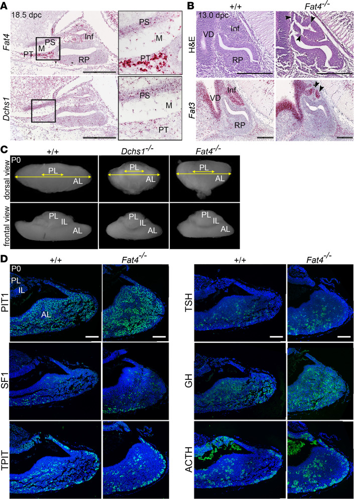
Figure 4. FAT4 and DCHS1 are required for normal murine pituitary development.
(A) RNAscope mRNA in situ hybridization on sagittal sections through WT murine pituitaries at 18.5 dpc using probes against Fat4 and Dchs1 (n = 3). Abundant Fat4 transcripts are detected in the pars tuberalis, infundibulum, developing pituitary stalk, and mesenchyme surrounding definitive RP. Some transcripts are also detected in RP. Expression of Dchs1 is detected at low levels throughout these tissues. (B) Hematoxylin and eosin staining of sagittal sections through Dchs1–/– (n = 5), Fat4–/– (n = 10), and control pituitaries (n = 15) at 13.0 dpc showing invaginations in the infundibulum of Fat4–/– mutants (arrowheads), not observed in control or Dchs1–/– embryos. RNAscope mRNA in situ hybridization on sagittal sections through control WT and Fat4–/– pituitaries at 13.5 dpc using specific probes against Fat3 marking the ventral diencephalon and infundibulum, which is abnormal in mutants (arrowheads). (C) Wholemount images taken at dorsal (top panels) and frontal views (bottom panels) of control, Dchs1–/– (n = 10), and Fat4–/– (n = 8) pituitaries at P0. Both Dchs1–/– and Fat4–/– mutants have a shortened medio-lateral axis affecting the anterior lobe compared with control. (D) Immunofluorescence staining on Fat4–/– pituitaries and littermate controls at 18.5 dpc using antibodies against lineage-committed progenitor markers PIT1, TPIT, and SF1 and hormones TSH, GH, and ACTH (n = 3). Staining is comparable for all markers between genotypes. Inf, infundibulum; PS, pituitary stalk; PT, pars tuberalis; M, mesenchyme; VD, ventral diencephalon; PL, posterior lobe; IL, intermediate lobe; AL, anterior lobe. Scale bars: 250 μm (A and B), 100 μm (D). For insets in A, original magnification, ×2.7.

