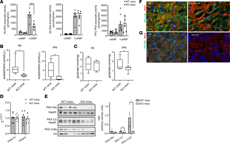
Figure 2. RIIα-KO mice had reduced habenular PKA enzymatic activity, decreased IPN acetylcholine and glutamate levels, and altered localization of PKA catalytic subunit to dendrites compared with WT mice.
(A) Basal and total (cAMP-stimulated) PKA enzymatic activities in Hb, striatum (Str), and prefrontal cortex (PFC); n = 5–8/group (female data shown). (B) Acetylcholine concentrations in Hb and IPN were lower in RIIα-KO mice compared with WT littermates; n = 6–7/sex/group (male data shown). (C) Glutamate concentrations did not differ in Hb but were lower in IPN of KO compared with WT mice; n = 7–9/group (female Hb and male IPN data shown). (D) Habenular Prkar1a and Prkaca mRNA levels did not differ between WT and RIIα-KO mice; n = 7/group (male data shown). (E) Representative Western blots of Hb lysates for PKA subunits RIα and Cα (first membrane with Gapdh as housekeeper, females) and combined Cαβγ (second membrane with Histone 3 as housekeeper, males); n = 4/genotype. Representative immunofluorescent images of WT (left) and RIIα-KO (right) brain sections (MHb) showed differences in the subcellular localization of (F) PKA catalytic subunits (αβγ, green) in lower dMHb in WT, and mutant mice that had impaired dendritic localization (shown by staining for MAP2, red), and (G) PKA RIIα (green) that is localized both to the cell body and dendrites (MAP2, red) in WT mice (female data shown). *P < 0.05; **P < 0.01, ***P < 0.001, unpaired 2-tailed t tests. All data represent the mean ± SEM.

