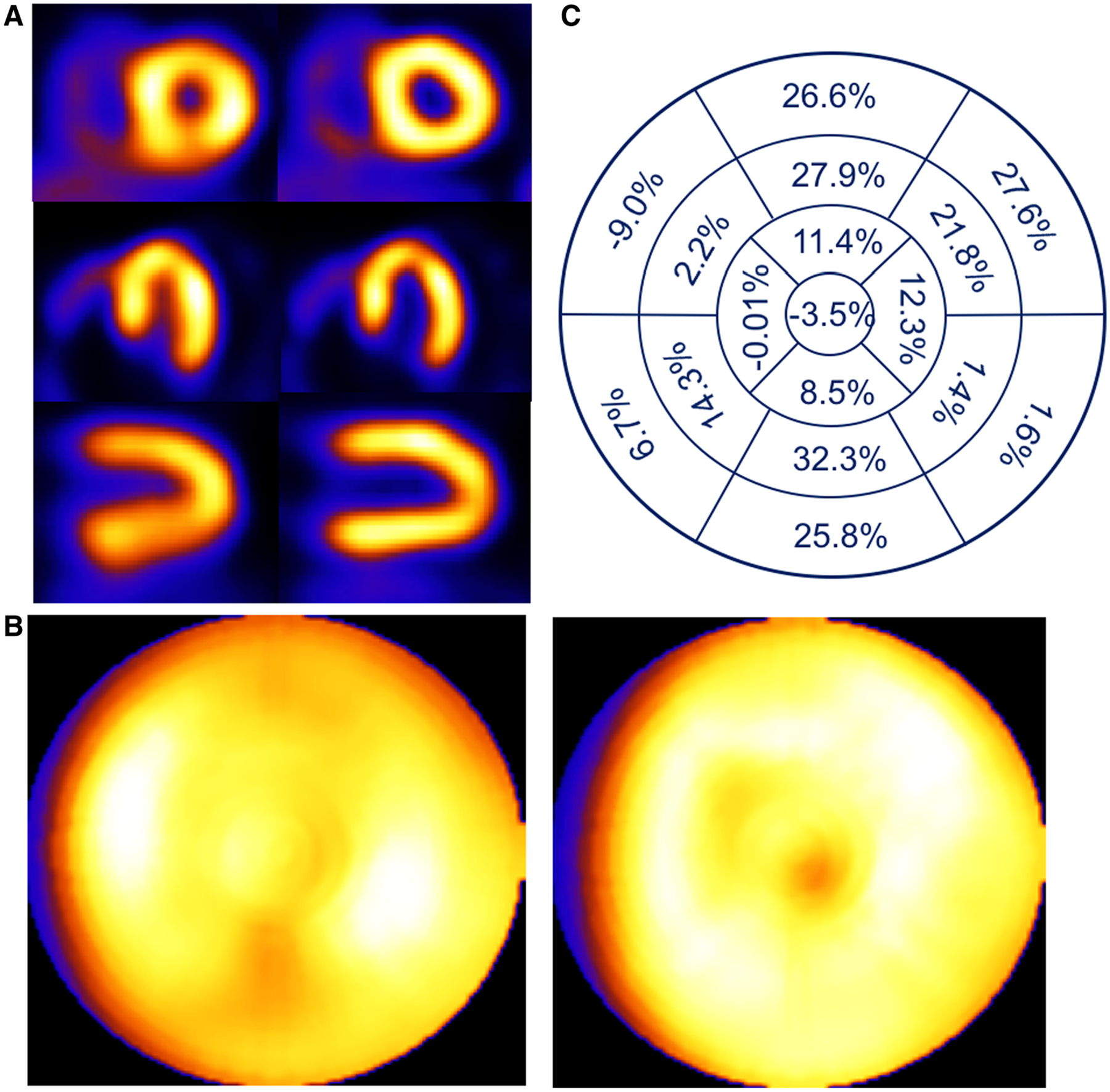
Figure 7.

Patient example of a 54-year-old female with BMI of 35.3 presenting with palpitations. Respiratory motion estimates were 2.9, 7.3, and 21 mm in the x (left-to-right), y (anterior-inferior), and z (head-to-foot) directions respectively. (A) Shown are short-, horizontal long-, and vertical long-axis slices without (left) and with (right) respiratory compensation. (B) Comparison of polar maps generated from short-axis slices without (left) and with (right) respiratory motion compensation. (C) Presentation of the average % segmental-count differences between without and with respiratory motion compensation for the 17 polar map segments.
