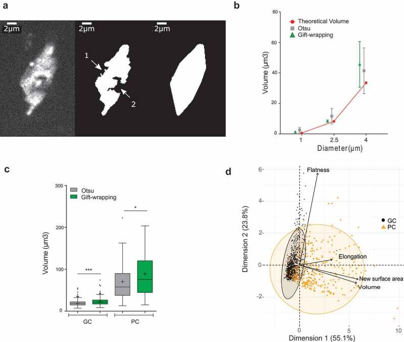
Figure 2.

Evaluation of a 3D gift-wrapping method of segmentation
(a) Example of a nucleus raw slice (left) after Otsu-modified segmentation (middle) and gift-wrapping segmentation (right). Artefactual indentation at the nuclear border (arrow #1); Nucleolus border (arrow #2).(b) Comparison of Otsu and gift–wrapping methods using standardized microspheres. Microsphere volume of 1, 2.5 and 4 µm diameter (n = 28, 24, and 15, respectively) were computed by the Otsu (green triangle) and gift-wrapping (gray square) methods and compared to theoretical volumes (red circle) (Supplemental table 2).(c) Comparison of nuclear volumes after segmentation of plant nuclei by the Otsu or gift-wrapping methods. Nuclei were split into two categories: guard cells (n = 375) and pavement cells (n = 127) (Supplemental table 4) were segmented by the two methods and volumes of the segmented nuclei were computed by NucleusJ 2.0. Modified Otsu method (gray); gift-wrapping (green). Student t-test P-value: *** <0.0001, * = 0.0046.(d) Principal component analysis of morphology parameters (Flatness, Elongation, New surface area and Volume) obtained after segmentation by the gift-wrapping (left) or Otsu (right) methods of the same nuclei as in Figure 2c. Guard cell nuclei (GC, black) and pavement cell nuclei (PC, orange).
