
Figure 1.
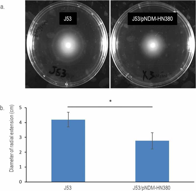
Motility of E. coli J53 and J53/pNDM-HN380. (a). Representative pictures of radial extension in 0.3% soft LB agar. Bacterial strains were labelled accordingly. (b). Diameters of the radial extension halo. The experiments were conducted in biological triplicates. Bars indicate the standard deviation. * p-Value <0.05