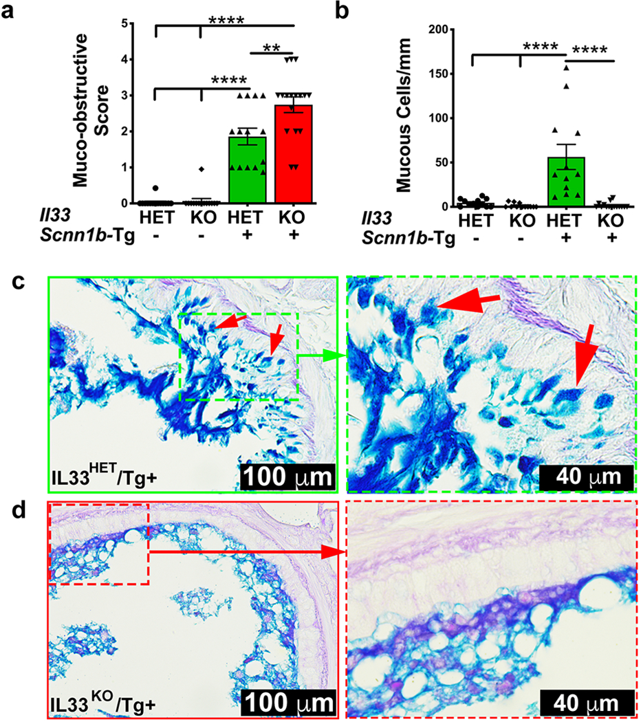
Figure 4: IL33 deletion does not ameliorate mucus obstruction despite significant suppression of mucous cell density in Scnn1b-Tg+ airways.

(a) Semi-quantitative histological scoring for airway mucus obstruction IL33HET/WT (white bar), IL33KO/WT (blue bar), IL33HET/Tg+ (green bar), IL33KO/Tg+ (red bar). (b) Number of mucous cells per mm of basement membrane, IL33HET/WT (white bar), IL33KO/WT (blue bar), IL33HET/Tg+ (green bar), IL33KO/Tg+ (red bar). (c) Representative photomicrographs from AB-PAS-stained left lung sections from IL33HET/Tg+ (higher magnification of inset is shown as dotted green margin). Intracellular AB-PAS staining of mucous cells is indicated by red arrows. (d) Representative photomicrographs from AB-PAS-stained left lung sections from IL33KO/Tg+ (higher magnification of inset is shown as dotted red margin). Error bars represent SEM. **p<0.01, ****p<0.0001 using ANOVA followed by Tukey’s multiple comparison post hoc test.
