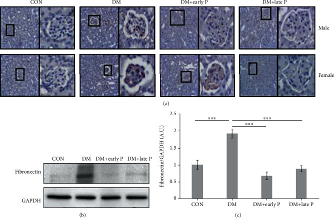
Figure 5.

Villin expression in mouse renal tissues. (a) Immunohistochemistry analysis of villin expression in mouse renal sections. Staining intensity of villin was lower in DM as compared to control (CON) mice. Paricalcitol treatment (DM+early P, DM+late P treatments) restored villin levels. Magnification, ×20. (b) Representative western blots of villin and GAPDH proteins extracted from mouse renal tissues. (c) Quantification of total western blots. Renal villin levels were significantly lower in DM mice (n = 12) as compared to control mice (n = 11) (∗p < 0.001) and were significantly higher in paricalcitol-treated mice (DM+early P (n = 10), DM+late P (n = 13) treatments) as compared to DM mice (∗∗p < 0.01, ∗∗∗p < 0.01, respectively). Error bars correspond to SEM.
