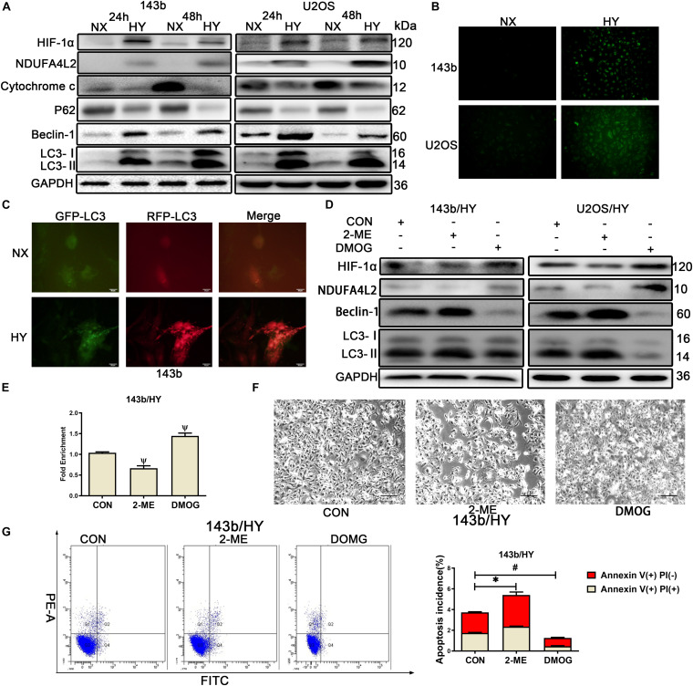
FIGURE 1.
HIF-1α might regulate the expression of NDUA4L2 in 143b and U2OS cell lines. 143b and U2OS cells were cultured in hypoxic environments for 24 and 48 h while control cells were cultured in normoxic environments. (A) Protein expression of HIF-1α, NDUFA4L2, P62, Beclin-2, LC3, and GAPDH in 143b and U2OS cells was determined by Western blotting. (B) ROS production was detected in 143b and U2OS under normoxic environments or hypoxic environments for 48 h by use of a Reactive Oxygen Detection Kit. (C) Fluorescence-based imaging for 143b cells transfected with mRFP-GFP-LC3 adenovirus. Green dot represents the start of autophagic flux, and Red dot represents the end of autophagic flux. Promotion of Red dot represents that autophagic flux was promoted. 143b cells were under normoxic environments or hypoxic environments for 48 h. (D) Expression of HIF-1α, NDUFA4L2, Beclin-1, LC3, and GAPDH were determined by Western blotting in 143b and U2OS cells pretreated with 2-ME and DMOG in hypoxic environments. (E) CHIP assay of HIF-1α binding to NDUFA4L2 gene in 143b cells. (F) About 1 × 10⁶ 143b cells were planted into six-well plates and cultured at 37•C until 70% confluence. 143b cell morphology was photographed using microscopy. Scale Bar = 100 μm. (G) Apoptosis of 143b cells was detected by flow cytometry. NX, normoxic environment; HY, hypoxic environment. nsp ≥ 0.05, ∗p < 0.05, ψ < 0.01, and # < 0.001 were defined as measures that indicated significant differences among treatment groups. All experiments were performed in triplicate.

