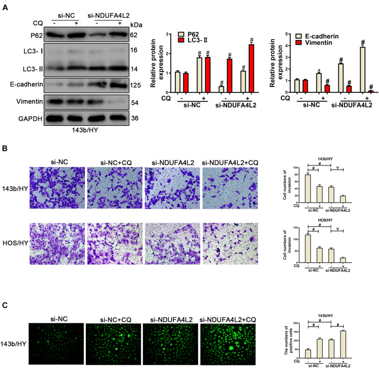
FIGURE 5.
Repression of autophagy could repress the EMT progression of si-NDUFA4L2-transfected 143b and HOS cells. 143b and HOS cells were treated with CQ after transfecting with si-NC or si-NDUFA4L2 in hypoxic environments. (A) Western blotting was performed to determine the protein expression of P62, LC3, E-cadherin, Vimentin, and GAPDH in 143b cells. For P62, LC3, E-cadherin, and Vimentin, si-NC vs. si-NC + Rapamycin was # < 0.001, si-NC vs. si-NDUFA4L2 was # < 0.001, si-NDUFA4L2 vs. si-NDUFA4L2 + Rapamycin was # < 0.001. (B) Transwell assays were performed to determine the invasion ability of si-NDUFA4L2-transfected 143b and HOS cells after treatment with CQ. (C) ROS production was detected by using Reactive Oxygen Detection Kits. HY, hypoxic environment. nsp ≥ 0.05, ψ < 0.01, and # < 0.001 were defined as measures that indicated significant differences among treatment groups. All experiments were performed in triplicate.

