Figure 6.

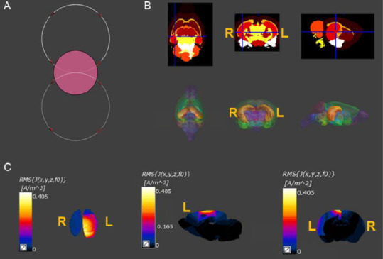
Finite element analysis of the magnetic stimulation-induced electrical field and magnetic field distribution in the 3D brain model of MCAO rats.
(A) Effects of different spatial positions of the figure-8 coil on the electromagnetic field distribution: The diameter of the circular coil and the single diameter of the figure-8 coil were both 12 mm. The coils were placed 2 mm above the plane of the head. (B) The 3D model included 154 brain regions, and the corresponding conductivity and permittivity according to the physical properties of the brain tissues. (C) The figure-8 coil simulation results of the 3D model of MCAO rats. Compared with the contralateral side, the distribution of the electromagnetic field within and around the liquefied necrotic area showed significantly enhanced current density. 3D: Three-dimensional; L: ipsilateral cortex; R: contralateral cortex.
