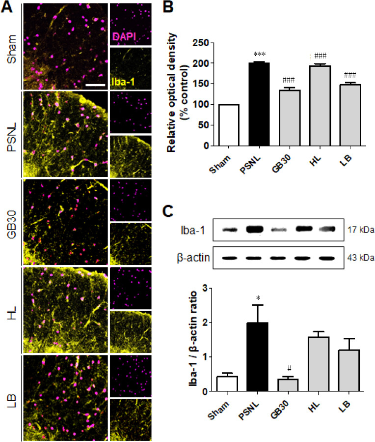
Figure 6.

Regulation of muscovite in activated microglia.
(A, B) Immunofluorescence staining demonstrates that Iba-1 is regulated for muscovite injection. (C) Western blot assay confirming Iba-1 expression after muscovite injection. β-Actin was used as the loading control. Scale bar: 100 µm. Data are expressed as the mean ± SEM (n = 3/group). *P < 0.05, ***P < 0.001, vs. sham group; #P < 0.05, ###P < 0.001, vs. PSNL group (one-way analysis of variance followed by the Student-Newman-Keuls test). DAPI: 4′,6-Diamidino-2-phenylindole; HL: hindlimb (BL25); LB: lumbar (GB34); PSNL: partial sciatic nerve ligation.
