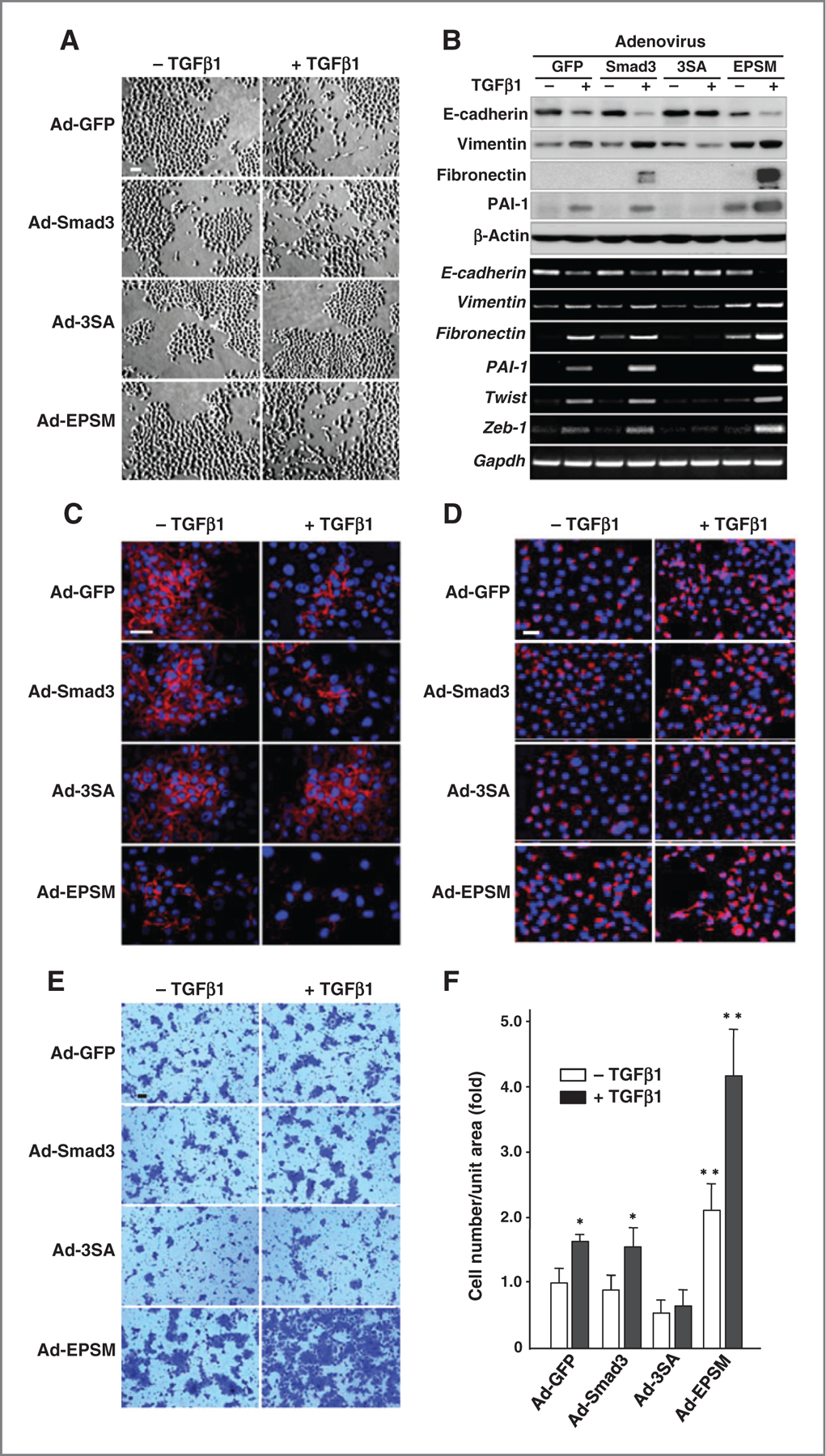
Figure 4.

Mutation of Smad3 linker phosphorylation sites accelerates the TGFβ-induced EMT together with the invasive activity. A, phase-contrast micrographs of CA1a cells infected with Ad-GFP, Ad-Smad3, Ad-3SA, or Ad-EPSM with or without TGFβ1. Scale bar, 50 µm. B, immunoblot analyses (top) and RT-PCR (bottom) for EMT markers (E-cadherin, vimentin, and fibronectin), PAI-1, and E-cadherin suppressors (Twist and Zeb-1) in the adenovirus-infected CA1a cells with or without TGFβ1. C and D, immunofluorescence staining of E-cadherin (red; C) and vimentin (red; D) counterstained with DAPI (blue). Scale bar, 50 µm. E, invading cells through Matrigel membrane. Scale bar, 50 µm. F, invasive activities (fold) calculated from results. The mean ± SD from four experiments. *, 0.01 < P < 0.05; ** P < 0.01, compared with Ad-Smad3–infected cells without TGFβ1. Each of the experiments was conducted three to five times with similar results.
