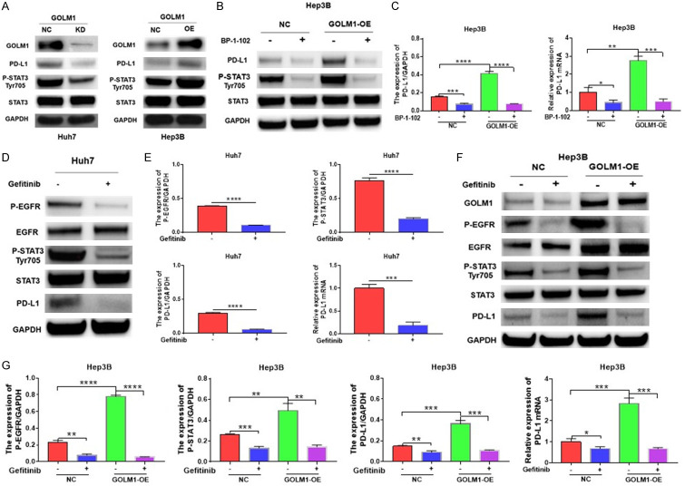
Figure 5.
GOLM1 promotes PD-L1 expression via the EGFR/STAT3 pathway. A. Western blot analysis of PD-L1 and phosphorylation of STAT3 Tyr705 in GOLM1-knockdown Huh7 cells and in GOLM1-overexpressed Hep3B cells. B. GOLM1 upregulates the expression of PD-L1 through promoting phosphorylation of STAT3 Tyr705 (P-STAT3). The expression of PD-L1, P-STAT3 and STAT3 was detected by Western blot after incubation with BP-1-102 (5 μM) for 12 hours in Hep3B cells. C. Quantitative analysis of PD-L1 expression after treatment with BP-1-102 through Image J intensity measurement. The relative expression of PD-L1 mRNA in Hep3B cells and GOLM1-overexpressed Hep3B cells was detected by RT-qPCR after incubation with BP-1-102 (5 μM) for 12 hours. D. The expression of EGFR, the phosphorylation of EGFR (P-EGFR), STAT3, P-STAT3 and PD-L1 was detected by Western blot after treatment with Gefitinib (4 μM) for 6 hours in Huh7 cells. E. Quantitative analysis of P-EGFR, P-STAT3 and PD-L1 expression after treatment with Gefitinib through Image J intensity measurement. The relative expression of PD-L1 mRNA was detected by RT-qPCR after treatment with Gefitinib in Huh7 cells. F. GOLM1 enhances the phosphorylation of STAT3 Tyr705 through the regulation of P-EGFR and EGFR. The expression of GOLM1, EGFR, P-EGFR, STAT3, and P-STAT3 was detected by Western blot in Hep3B cells and GOLM1-overexpressed Hep3B cells after incubation with Gefitinib. G. Quantitative analysis of P-EGFR, P-STAT3 and PD-L1 expression after treatment with Gefitinib through Image J intensity measurement. The relative expression of PD-L1 mRNA in Hep3B cells and GOLM1-overexpressed Hep3B cells was detected by RT-qPCR after treatment with Gefitinib. Cells were incubated with EGF (50 ng/ml).

