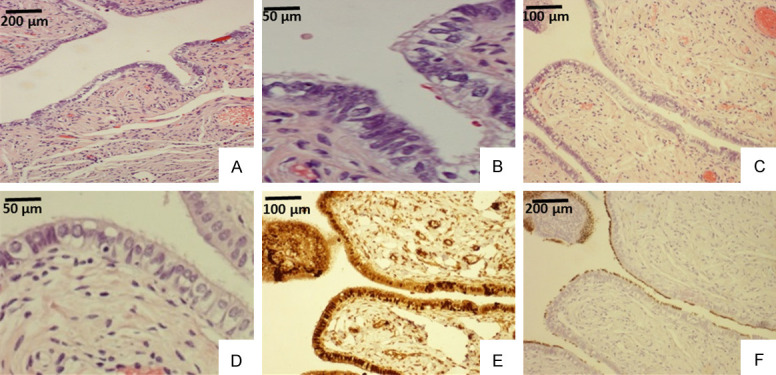
Figure 5.

Example of tubal ciliated cells detected by microscopy and tubulin staining in a patient of 45 years old. Tubal ampulla segment (A, B) showed ciliated cells, which are easily visible in a high power (upper right corner of B). Tubal fimbria region (C, D) showed cilia on the apical cellular border under a high power view (D). The same fimbria region (C) stained with PAX8 for tubal secretory cells (E) and tubulin for ciliated cells (F). Apparently, PAX8 stained both secretory and ciliated cells (E) as the ciliated cells were illustrated by tubulin stain (F). Original magnifications: (A, C, E, and F) 100×; (B and D), 400×.
