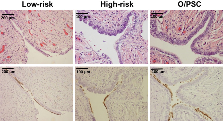
Figure 6.

Morphologic and immunohistochemical identification of tubal ciliated cells in tubal fimbria. One representative section of tubal fimbria from patients in an age group of 40s was presented. Top panel shows morphologic picture of tubal fimbria, while bottom panel shows corresponding tubulin stains. The number of ciliated cells (tubulin+) was 180, 128, and 141 for low-risk, high-risk, and O/PSC patients, respectively. Original magnifications: left 100×, middle 200×, right 200×.
