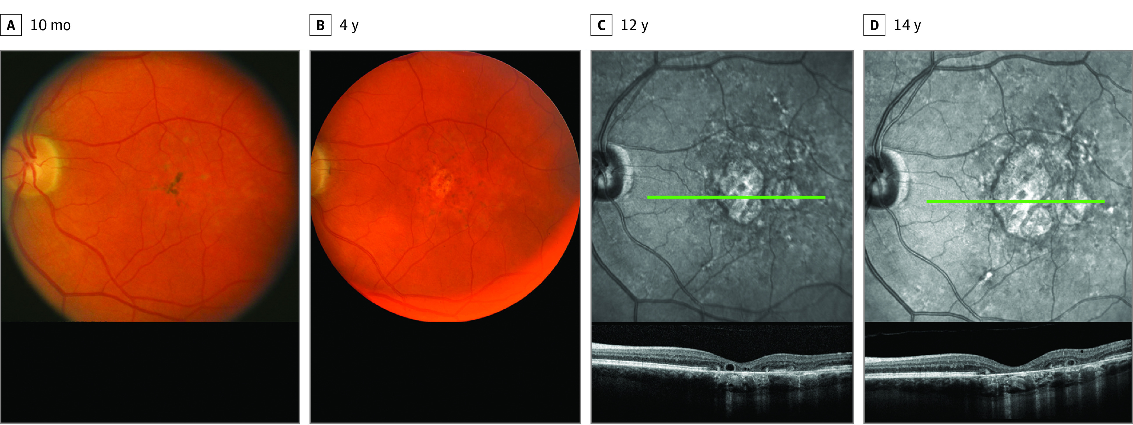
Figure 2. Maculopathy Progression After Completion of Blood-Brain Barrier Disruption Therapy in a Patient Treated for a Central Nervous System Glioma.

A and B, Progression of maculopathy shown by fundus photography at 10 months and 4 years. C and D, Near-infrared images in the same patient with corresponding optical coherence tomography (OCT) demonstrates expanding complete retinal pigment epithelium and outer retinal atrophy, as well as outer retinal tubulations (green lines designate the location of corresponding B-scan OCT images below) at 12 and 14 years.
