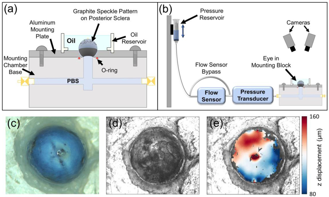
Fig. 2.
Whole-globe inflation testing setup. a Schematic of the testing rig. b Schematic of the whole globe inflation testing apparatus. c Overhead view of the sclera before graphite speckle pattern was applied. d DIC camera view of the sclera after application of graphite speckle pattern. e Facet overlay displaying z displacement (toward the camera) of the scleral surface at the end of an inflation test. a and b from (Hannon et al. 2019)

